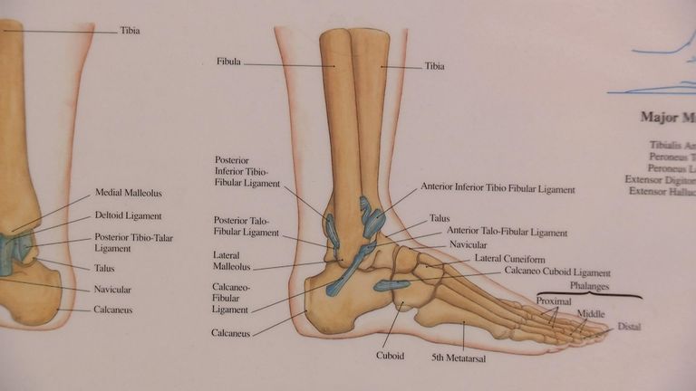
Bones In Leg Diagram. The bones of the leg are the femur, tibia, fibula and patella. Normal leg bones are relatively straight, but those affected by paget's disease are porous and curved. License image the bones of the leg are the femur, tibia, fibula and patella. The foot bones shown in this diagram are the talus, navicular, cuneiform, cuboid, metatarsals and calcaneus. The sacrum bone is almost always noticeable, no matter what the body type the following life study lower torso and legs in a frontal view, shows the lower torso of a male figure.
Want to learn more about it? Leg diagram illustrations & vectors. The thigh bone (femur) is the longest bone in the body. The femur, or thigh bone, is the largest, heaviest, and strongest bone in the human body. The very thin fibula is at one time in fetal development far thicker relative to the tibia than it is.
Luke Shaw expected to be out for up to nine months, expert ... from e2.365dm.com These leg muscle diagrams show you the major muscles of the human leg. When you stand or walk, all the weight of your upper body rests on them. Your leg bones are very large and strong to help support the weight of your body. Click and start learning now! The femur, or thigh bone, is the largest, heaviest, and strongest bone in the human body. At the distal end of the femur, two rounded condyles meet the tibia and fibula bones of the lower leg to form the knee joint. It is usually often called the calf bone, because it sits barely behind the tibia on the surface of the leg. It joins with humerus on its larger end to make elbow joint and join with the carpal bone of the hand at its smaller end.
There are exactly 26 bones in the hand and 26 in the foot.
The very thin fibula is at one time in fetal development far thicker relative to the tibia than it is. 12 photos of the bones leg diagram picture. These leg muscle diagrams show you the major muscles of the human leg. Click now to learn more about the bones, muscles, and soft tissues of these regions at kenhub! Click and start learning now! Leg diagram illustrations & vectors. He leg's main function in the human is for locomotion and support of the rest of the body. Schema de legs bones diagram diagram showing bones inside human leg ready to jump stock file skeleton of a cat diagram ver 2 svg disposition of rotator cuff muscles diagram. The majority of muscles in the leg are considered long muscles, in that they stretch great distances. It joins with humerus on its larger end to make elbow joint and join with the carpal bone of the hand at its smaller end. Explore more like human leg bones diagram. An electrical wiring diagram can be as simple as a diagram demonstrating how to set up a fresh swap with your hallway. The thigh bone (femur) is the longest bone in the body.
Nervsystemet anatomy, diagram & function | health. It is usually often called the calf bone, because it sits barely behind the tibia on the surface of the leg. Editor · aug 13, 2017 ·. Also, defective and old red blood cells are destroyed in bone marrow. These leg muscle diagrams show you the major muscles of the human leg.
Biology, Animal Structure and Function, The ... from oercommons.s3.amazonaws.com Leg muscle sport trauma and bone pain labeled diagram. Top suggestions for human leg bones diagram. The foot bones shown in this diagram are the talus, navicular, cuneiform, cuboid, metatarsals and calcaneus. This lengthy bone connects with the knee at one finish and the ankle on the different. Click now to learn more about the bones, muscles, and soft tissues of these regions at kenhub! The thigh bone (femur) is the longest bone in the body. We discuss their function, the different types of bones in the human body, and the cells that are involved. Cancellous bone produces red blood cells, platelets, and white blood cells.
Related posts of leg bones anatomy diagram structure of anatomy leg and foot.
Editor · aug 13, 2017 ·. The femur (thigh bone), tibia and fibula (lower leg bones), clavicle (collar. The foot bones shown in this diagram are the talus, navicular, cuneiform, cuboid, metatarsals and calcaneus. Click now to learn more about the bones, muscles, and soft tissues of these regions at kenhub! The bones involved in it, however, are only the femur and the tibia, although the smaller bone of the leg, the fibula, is carried along in the movements of flexion, extension, and slight rotation that this joint permits. Bones pain hand and arm bones diagram. The majority of muscles in the leg are considered long muscles, in that they stretch great distances. Continue scrolling to read more below. Related posts of leg bones anatomy diagram structure of anatomy leg and foot. The bone that goes from your pelvis to your knee is called the femur (say: The knee is a strong but flexible hinge joint. Leg muscle sport trauma and bone pain labeled diagram. Learn vocabulary, terms and more with flashcards, games and other study tools.
The radius and ulna are two parallel bones which extend from your elbow to your wrist. Master leg and knee anatomy using our topic page. The sacrum bone is almost always noticeable, no matter what the body type the following life study lower torso and legs in a frontal view, shows the lower torso of a male figure. Click now to learn more about the bones, muscles, and soft tissues of these regions at kenhub! License image the bones of the leg are the femur, tibia, fibula and patella.
{Lower Leg Bones Pelvic-Femur-Tibia-Fibula-Foot} | John ... from www.johnthebodyman.com The knee joint is the largest joint in the body and is primarily a hinge joint, although. The bones of the leg are the femur, tibia, fibula and patella. When you stand or walk, all the weight of your upper body rests on them. Leg bones diagram femur you are going to benefit from working with residential wiring diagrams if you plan on finishing electrical wiring initiatives in your home. Also, defective and old red blood cells are destroyed in bone marrow. There are exactly 26 bones in the hand and 26 in the foot. Posted on january 20, 2015 by admin. The bones of the leg are the femur, tibia, fibula and patella.
Master leg and knee anatomy using our topic page.
When you stand or walk, all the weight of your upper body rests on them. Leg muscle sport trauma and bone pain labeled diagram. An electrical wiring diagram can be as simple as a diagram demonstrating how to set up a fresh swap with your hallway. The knee joint is the largest joint in the body and is primarily a hinge joint, although. Your leg bones are very large and strong to help support the weight of your body. Related posts of leg bones anatomy diagram structure of anatomy leg and foot. Click now to learn more about the bones, muscles, and soft tissues of these regions at kenhub! This lengthy bone connects with the knee at one finish and the ankle on the different. Cancellous bone produces red blood cells, platelets, and white blood cells. Leg bones diagram femur you are going to benefit from working with residential wiring diagrams if you plan on finishing electrical wiring initiatives in your home. Top suggestions for human leg bones diagram. These leg muscle diagrams show you the major muscles of the human leg. The bones of the leg are the femur, tibia, fibula and patella.


Social Media